Arterial Supply to the Structures of Balance
Posterior Circulation 1
The anterior structures of the brain are supplied by the carotid arteries, and will not be covered here. More appropriately for a balance and dizziness discussion, this page focuses on the posterior structures of the central nervous system and the posterior circulation. The posterior circulation, or vertebral-basilar system, traditionally begins with the right and left vertebral arteries.
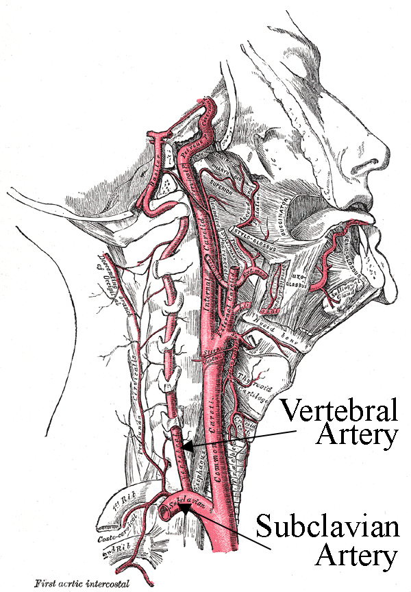
FIGURE 1
Wikimedia Commons
These are the first branches of the subclavian arteries (see figure 1). Subclavian simply refers to the arteries' position beneath/behind the clavicle ("collar bone"). Although not classically considered part of the vertebral-basilar system, it is important to note these vessels because of a germaine condition discussed later called the "Subclavian Steal Syndrome."
The vertebral arteries ascend via the cervical vertebrae and enter the cranium through the foramen magnum (literally the "big hole" where the spinal cord enters the skull). Just after entering the cranial space, the right and left vertebral arteries give rise to the right and left posterior spinal arteries, which travel down the posterior spinal cord (not pictured in figure 2).
Approximately 1-2cm before the vertebral arteries join they give rise to the posterior inferior cerebellar arteries (PICA). The bottom of figure 2 shows the right and left PICA branching from the right and left vetebral arteries. The PICA play a significant role in balance by supplying the lateral medulla (where the vestibular nuclei reside) and the entire posterior and inferior parts of the cerebellum (via the inferior vermian and tonsillar-hemispheric branches).
Roughly less than 1cm before joining, the vertebral arteries give rise to a final pair of vessels: the anterior spinal arteries. These two arteries join and descend along the anterior spinal cord.
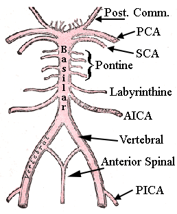
The vertebral arteries finally join near the pontomedullary junction (where the rostral medulla and caudal pons meet) to form the single basilar artery. The right and left anterior inferior cerebellar arteries (AICA) arise from the basilar artery near its origin. They project along the path of CN VII and CN VIII supplying the lateral pons and finally reaching their largest region of supply in the anterior and inferior parts of the cerebellum. The labyrinthine artery, the sole blood supply to the inner ear, either branches off from the AICA (most common) or from the basilar artery directly.
The pontine arteries branch from the middle of the basilar artery to supply the medial pons. These are several small vessels rostral to the labyrinthine arteries. Just before the basilar artery divides at the midbrain, it gives rise to right and left superior cerebellar arteries (SCA). These divide into the hemispheric and superior vermian branches, which supply the superior portion of the cerebellum, most of the cerebellar nuclei, and the superior cerebellar peduncles.
Upon reaching the midbrain the basilar artery bifurcates into right and left posterior cerebral arteries (PCA). The PCA anastomose with the posterior communicating arteries to connect the posterior and anterior circulations in the circle of Willis. While the PCA also supplies several areas, specifically to balance and dizziness, the PCA provides blood to the midbrain through several branches (thalamoperforate, medial posterior choroidal and quadrigeminal).
This may all sound a little overwhelming, but use figure 2 to observe how most of these arteries are merely branching from the central vertebral-basilar arteries (hence the name of the system). Now test your mettle and see if you can identify the arteries of the posterior circulation by taking the short quiz! Compare the quiz illustration to the labels in figure 2 for help.

Labyrinth of the Inner Ear 2
As mentioned above, a single artery, the labyrinthine/internal auditory artery, supplies the inner ear with oxygenated blood. It most often rises from the AICA, but in some cases stems directly from the basilar artery. The labyrinthine artery is an end artery, thus does not communicate with any other vessel in the temporal bone. Because of the inner ear's singular blood supply, both the vestibular and auditory structures are especially vulnerable to ischemic events. Below is a flow chart depicting the rather complex arterial supply to the vestibular apparatus and cochlea.

The labyrinthine artery has two main branches: the common cochlear and the anterior vestibular arteries. The common cochlear artery gives rise to the main cochlear artery, which radiates from the modiolus to supply the spiral ganglion, organ of Corti and striavascularis. The posterior vestibular artery, another branch of the common cochlear artery, supplies the majority of the saccule (inferior part) and the ampulla of the posterior semicircular canal.
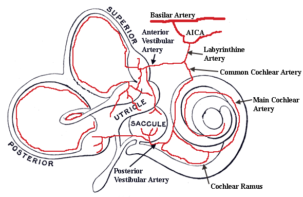
The second branch of the labyrinthine artery is the anterior vestibular artery. This vessel supplies the ampullae of the anterior and horizontal semicircular canals, the utricle, and a small portion of the saccule (superior portion).
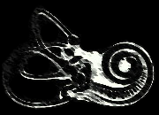
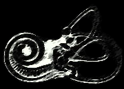
The branches of the labyrinthine artery are independent, which means occlusion of one branch only affects the structures supplied by that branch. For example, occlusion of the anterior vestibular artery does not lead to hearing loss. The signs and symptoms due to occlusion of the labyrinthine vasculature are discussed in detail in the following section. Below is an illustration showing the approximate course of the labyrinthine blood supply.


- Page References - Click the links below for full citations
- Basic Clinical Neuroscience
- Clinical Neurophysiology of the Vestibular System (Third Edition)
- MedlinePlus Medical Dictionary

