The Medulla Oblongata (Myelencephalon)
In essence, the brain stem connects the cerebrum to the rest of the body; it does this through joining the brain to the cerebellum and spinal cord. The most caudal structure, communicating directly with the spinal cord, is the medulla oblongata.
Anatomy of the Medulla
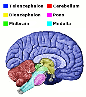
FIGURE 1 Wikimedia Commons
Cranial Nerves 1 2
Each of the three parts of the brainstem serve as points of origin to specific cranial nerves (CN). (A detailed treatment of the 12 cranial nerves can be found in their dedicated section.) Learning which cranial nerves are associated with each structure of the brainstem is paramount in determining the general site of lesion. This knowledge will help you predict and make sense of test results, as well as indicate when your test results may need to be repeated (i.e., your results do not make sense); more on this below.
The cranial nerves associated with the medulla are:
- CN IX - the glossopharyngeal nerve
- CN X - the vagus nerve
- CN XI - the spinal accessory nerve
- CN XII - the hypoglossal nerve
As a brief review, CN IX (glossopharyngeal) is important for swallowing, talking and taste; CN X (vagus) is also involved with the muscles of swallowing and speech, as well as slowing the heart rate and constriction of smooth visceral muscle; CN XI (spinal accessory) innervates the sternocleidomastoid and trapezius muscles (neck and back, respectively); and CN XII (hypoglossal) innervates all of the muscles of the tongue except for one.
Blood Supply1 2
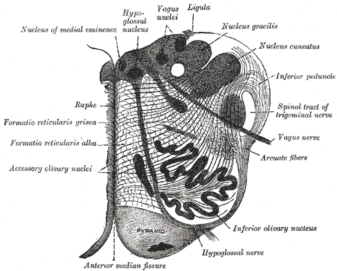
FIGURE 2 Wikimedia Commons
The medulla oblongata receives oxygenated blood from the branches of the vertebral and basilar arteries. For detailed coverage of these vessels and the rest of the posterior circulation, visit the vascular system section. The medulla can be divided into three main "zones" of irrigation, and because these zones contain specific structures and receive blood from specific vessels, becoming familiar with these areas will greatly assist you in identifying which blood vessels/medullary structures might be affected by a vascular event.
The Anterior Zone. The main structures located in the anterior zone are the pyramids, the medial longitudinal fasciculus (MLF), internal arcuate fibers and/or medial lemniscus, and CN XII.1 The caudal blood supply (near the spinal cord) comes from the anterior spinal artery. The rostral supply (upper medulla) is from the vertebral and basilar arteries.
The Lateral Zone. This zone contains "the nucleus ambiguous of CN X, the exiting fibers of CN IX and X, portions of the spinal nucleus and tract of CN V, ventral part of the inferior cerebellar peduncle, spinothalamic tracts, descending autonomics and the inferior olivary nucleus."1 The lateral zone is supplied caudally via the vertebral arteries and posterior inferior cerebellar artery (PICA). Rostrally, blood flow comes from the basilar artery and anterior inferior cerebellar artery (AICA).
The Posterior Zone. The structures of the posterior zone include: "the posterior white columns and their nuclei (gracilis and cuneatus), the caudal part of the vestibular nuclei, the dorsal motor nucleus of CN X, the nucleus and tractus solitarius, the inferior cerebellar peduncle and the spinal nucleus and tract of CN V."1 The majority of blood supply to the posterior zone is from the posterior spinal arteries.

Function of the Medulla
Further independent study of the particular structures associated with the various medullary zones will reveal the functions of the medulla in detail. Some of these structures are covered in this website, though many are not. In an effort to keep this section as focused on balance as possible, the following is a general synopsis of the roles of the medulla oblongata. Remember that mastering the anatomy of the medulla will lead naturally to its functions, as well as signs and symptoms associated with medullary syndromes.
- Functions of the Medulla Oblongata
- Speech and swallowing
- motor reflex control of larynx, pharynx and tongue
- Coughing, salivating, vomiting, sneezing and taste
- motor control of visceral reflexes
- Coordination of eye movements and positioning of the head and neck
- medial longitudinal fasciculus (MLF)
- Relay for cochlear and vestibular signals
- CN VIII
- Regulation of consciousness, visceral functions, sensation, etc.
- reticular formation


Lesions of the Medulla
Vascular Lesions1 2
Familiarity with the vessels, their regions of supply, and the structures of the medulla aids in determining the site of lesion. For example, should the lesion be vascular in origin, accompanying signs and symptoms will correspond to which tissues are damaged along an occluded vessel's supply. For a more in-depth discussion of the disruption of blood flow, please visit that section. What follows below is a short description of common medullary stroke syndromes.
| Syndrome | Description |
|---|---|
| Lateral Medullary Syndrome (Wallenberg Syndrome) | Click the name for a detailed review of etiology, signs and symptoms of this syndrome in the vascular system section of this website. |
| Medial Medullary Syndrome (Inferior Alternating Hemiplegia) | Unilateral spastic paralysis of the arm and leg caused by lesion of the corticospinal tract (this occurs on the contralesional side). Unilateral loss of proprioception (same side as paralysis) in the lower extremity due to involvement of the medial lemniscus (this occurs on the contralesional side). Atrophic tongue (tongue points to side of lesion when it is protruded) due to muscle weakness from lesion of CN XII (this occurs on the ipsilesional side). |
Neoplasms1 2
| Neoplasm | Description |
|---|---|
| Medulloblastoma | These tumors are most common in children, and can cause headaches, nystagmus and vomitting, stumbling gate, and diplopia. Headache is due to the tumor partially blocking the circulation of cerebrospinal fluid, which leads to buildup of fluid and intracranial pressure. Nystagmus and vomitting are elicited by involvement of the floor of the 4th ventricle (vestibular and vagal nerve nuclei). Broad-based gait with truncal ataxia is a sign of cerebellar involvement - specifically damage to the midline cerebellum. Double vision can be due to lesion of the MLF and/or abducens nuclei. |


- Page References - Click the links below for full citations
- Neuroanatomy Lab Resource Appendices
- Basic Clinical Neuroscience
- Isolated acute vertigo in the elderly; vestibular or vascular disease?
- MedlinePlus Medical Dictionary
- Inner Ear Dysfunction Due to Vertebrobasilar Ischemic Stroke
- Long-term observations on the reversibility of cochlear dysfunction after transient ischemia.
- Clinical Neurophysiology of the Vestibular System (Third Edition)
- NINDS Wallenberg's Syndrome Information Page
- Roles of the Cerebellum in Motor Control

